


Partial tasks
> Radiation Imaging in Material Science
Radiation Imaging in Material Science
Method “X Ray Dynamic Defectoscopy (XRDD)” uses Medipix device for the observation of time evolution of damage processes in a loaded specimen body. Even though the specimen is loaded monotonously and slowly, damaging can proceed discontinuously and relatively fast. Therefore high frame rate acquisition of X-ray images is crucial for real dynamic (time dependent) defectoscopy.
The method enables the observation of material density variations with micrometric accuracy. The test sample is illuminated by X rays during the loading process. Measured changes in transmission represent effective alterations in the sample thicknesses which are understood as weakening of the material due to damage volume fraction.
Sensitivity of the XRDD method depends on the ratio between incident and transmitted X ray beam intensity and on the fraction of scattered photons, the efficiency of the Medipix2 device at the X-ray energy used and the spectra of the X-ray tube.
Responsible person
Co-workers
Grants:
| Number | Name | Agency |
| LC06041 | Research center "Fabrication, modification and characterization of materials by energetic radiation" | MŠMT |
| 106/04/0567 | Study of damage zone in high ductile materials in vicinity of crack tip by X-Ray Dynamic Defectoscopy method | GAČR |
| MSM 6840770040 | Research Program 40: Usage of radionuclides and ionizing radiation. | MŠMT |
Articles in Impacted Journals
(21)
(21)
Text format

 Year
YearSearch
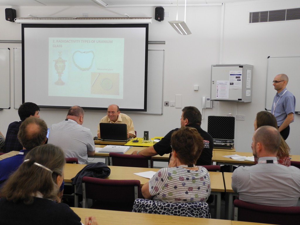
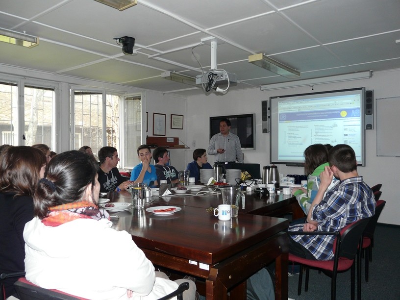
Recent events
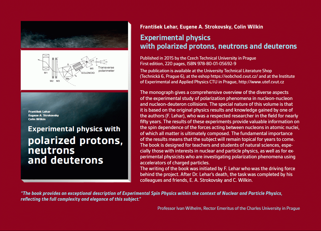 Experimental physics
with polarized protons, neutrons and deuterons
Experimental physics
with polarized protons, neutrons and deuterons Progressive detection methods in atomic and particle physics education at middle and high school level
 NSS MIC IEEE Conference
Seattle, USA
8-15 Nov 2014
 SEPnet, CERN@school Conference
Surrey, United Kingdom
Sep. 8, 2014
 Lovci záhad - natáčení ČT ve spolupráci s ÚTEF
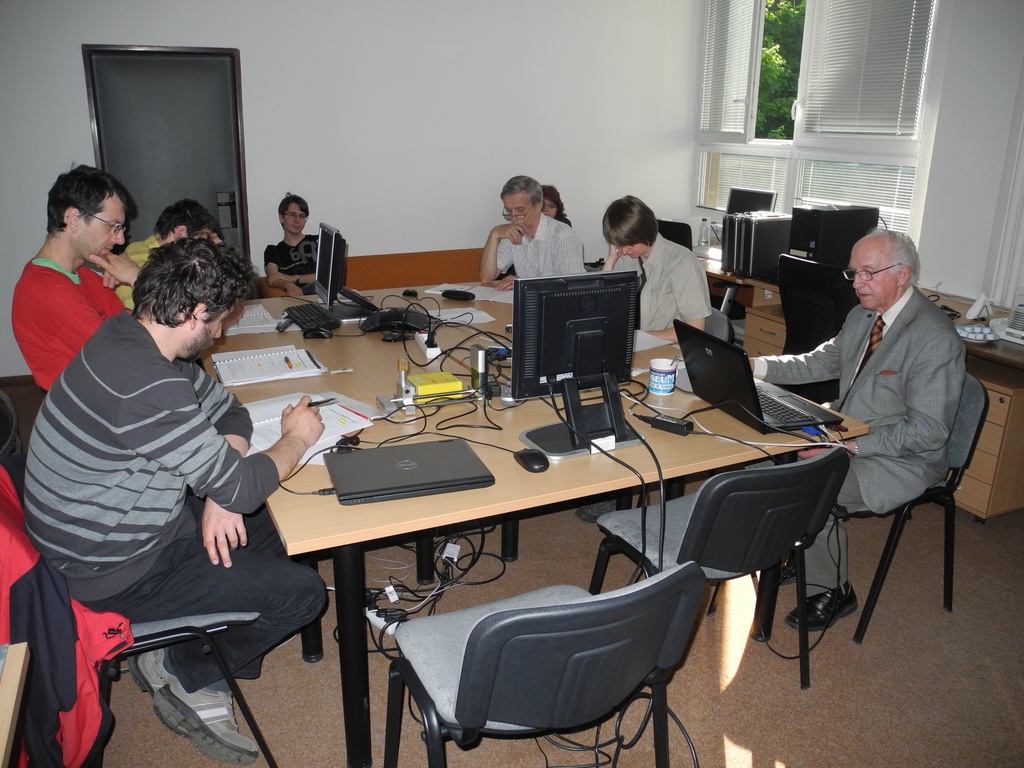 Advanced detection methods in atomic and subatomic physics education.
April 24, 2014
 Listening to the universe by detection cosmic rays - visit of French and Czech students
3 Apr 2014
Seoul, Korea
27 Oct - 2 Nov 2013
 15thIWORID
Paris
23-27 June 2013
 NSS MIC IEEE Conference
29 Oct - 3 Nov 2012
