


Publikace
> Příspěvky ve sbornících konferencí
> 'Real-Time X-Ray 2-D and 3-D Micro-Imaging of
Living Animals with Medipix2 Single Photon
Counting Detector'
Real-Time X-Ray 2-D and 3-D Micro-Imaging of
Living Animals with Medipix2 Single Photon
Counting Detector
Autor
| Frallicciardi Paola Maria | Dipartimento di Scienze Fisiche, Universita’ degli Studi di Napoli ”Federico II”, Napoli, Italy |
| Dammer Jiří, Mgr. | UTEF |
| Weyda František, doc. RNDr. CSc. | Biologické centrum Akademie věd České republiky, v. v. i. |
| Jakůbek Jan, Ing. Ph.D. | UTEF |
| Vavřík Daniel, Ing., Ph.D. | UTEF |
| Pospíšil Stanislav, Ing. DrSc. | UTEF |
Rok
2008
Časopis
Conf. Proc. IEEE NSS/MIC 2008, M10-112
Web
Obsah
In this work we present the study of applicability of a
desktop size radiographic/tomographic X-ray system for real-time
microscopy and micro-tomography in the fields of biology,
entomology, botanic and medical imaging. The apparatus is made
up of the single photon counting pixel silicon detector, Medipix2
(matrix of 256x256 square pixels of 55 μm pitch) and a microfocus
X-ray tube with a minimum spot size of 5 μm and a
tungsten anode. The system has been used for observations of
time-dependent processes inside living and still biological and
organic samples. Excellent contrast and spatial resolution
(micrometer scale) were obtained as a combination of a) low
photon energy (40 kVp X-ray tube voltage), b) single photon
counting operation, witch avoids integration of dark current c)
energy discrimination in each pixel, allowing noise rejection and
providing high SNR, d) high effective dynamic range for long
exposures, which allows for high signal with high SNR, e)
implementation of an original procedure for the energy response
calibration of each pixel of the detector matrix, f) high speed
read-out hardware and software, which opens the possibility to
perform real-time studies of biological processes permitting, e.g.,
observation of morphological changes, mutations or
metamorphosis of living animals and plants. Static and dynamic
images of a parasite life cycle from the larva stage to pupa stage are presented here, as well as an in vivo computed tomography of
the parasite living inside its host.
Granty
Projekty
Příklad citace článku:
P. Frallicciardi, J. Dammer, F. Weyda, J. Jakůbek, D. Vavřík, S. Pospíšil, "Real-Time X-Ray 2-D and 3-D Micro-Imaging of
Living Animals with Medipix2 Single Photon
Counting Detector", Conf. Proc. IEEE NSS/MIC 2008, M10-112 (2008)
Hledat
Události
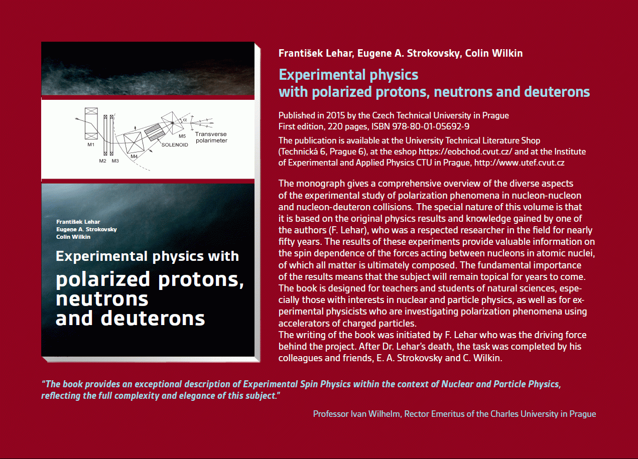 Experimental physics
with polarized protons, neutrons and deuterons
Experimental physics
with polarized protons, neutrons and deuterons Progresivní detekční metody ve výuce subatomové a částicové fyziky
na ZŠ a SŠ
Progresivní detekční metody ve výuce subatomové a částicové fyziky
na ZŠ a SŠ21.-22. 11. 2014
 NSS MIC IEEE Conference
Seattle, USA
8-15 Nov 2014
 Konference SEPnet, CERN@school
Surrey, Velká Británie
8. září 2014
 Lovci záhad - spolupráce ČT a ÚTEF
9. září 2014
 Progresivní detekční metody ve výuce subatomové a částicové fyziky na ZŠ a SŠ
24. 4. 2014
 Návštěva v rámci projektu „Listening to the universe by detection cosmic rays“
3. 4. 2014
Seoul, Korea
27 Oct - 2 Nov 2013
 15thIWORID
Paris
23-27 June 2013
 NSS MIC IEEE Conference
Anaheim, USA
29 Oct - 3 Nov 2012
