Publication
> Thesis
> 'X-Ray Phase Contrast Imaging with Hybrid Semiconductor Pixel Detectors'
X-Ray Phase Contrast Imaging with Hybrid Semiconductor Pixel Detectors
Author
Year
2014
Scientific journal
Ph.D. Thesis, Faculty of Biomedical Engineering, CTU in Prague
Web
Abstract
X-ray absorption imaging is currently a widespread imaging technique for nondestructive testing in industry as well as a main diagnostic tool for imaging of inner structures of the human body in medicine. In many situations, there is, however, the need to distinguish between low absorption and even low contrast features such as different kinds of soft tissue. In this case, the applicability of conventional absorption imaging can be limited. X-ray phase contrast imaging (XPCI) ¬is a very promising imaging technique providing enhanced contrast for certain combination of materials such as composite materials or different kinds of soft tissue with additional capability to reduce the radiation dose.
At present, the largest potential by the XPCI methods enabling high-quality imaging in a table-top setup is the grating based approach. During the last years it has been demonstrated that phase contrast imaging with a grating interferometer can be efficiently performed with a conventional, low-brilliance X-ray source with an enormous potential for applications in biology, medicine, nondestructive testing etc. However, to retrieve the phase gradient image is necessary to use so called phase-stepping approach. During phase-stepping, the grating is scanned transversely to the incident beam while acquiring multiple projections. The sample is supposed to be static, and the resulting poor time resolution is one of the major drawbacks of this method. Another critical fact is that phase stepping necessarily implies multiple exposures and, even though such exposures might be acquired each at very low dose, in comparison with conventional absorption imaging, the dose tends to be too high.
In this work, an alternative approach, which extracts the phase information without the need of a stepping procedure, thus overcoming limitations of both data acquisition speed and dose imparted to the specimen is introduced. The method is based on precise sub-pixel position determination of the X-ray pattern projected by the single absorption grating directly from the pattern image made possible by the application of highly sensitive hybrid pixel semiconductor detectors. The experimental results on a simple testing object as well as on complex biological samples are presented. The performance of the newly developed method is investigated to various setup parameters such as used X-ray spectrum and detector settings.
The last part of the Thesis is dedicated to the extensions of the Timepix detector technology to the extreme ultraviolet (XUV) spectral range which is for these devices in their original design intrinsically inaccessible. The proposed approach is experimentally verified in combination with a laser-generated plasma source of XUV in the ‘water window’ spectral range. The experimental results demonstrate high-sensitivity single-shot absorption radiography as a basic step towards development of advanced imaging techniques utilizing appropriate X-ray optics and also further contrast mechanism such as phase changes or scattering in this spectral range.
Projects
Cite article as:
F. Krejčí, "X-Ray Phase Contrast Imaging with Hybrid Semiconductor Pixel Detectors", Ph.D. Thesis, Faculty of Biomedical Engineering, CTU in Prague (2014)
Search
Recent events
Seattle, USA
8-15 Nov 2014
Surrey, United Kingdom
Sep. 8, 2014
April 24, 2014
3 Apr 2014
Seoul, Korea
27 Oct - 2 Nov 2013
Paris
23-27 June 2013
29 Oct - 3 Nov 2012


 Experimental physics
with polarized protons, neutrons and deuterons
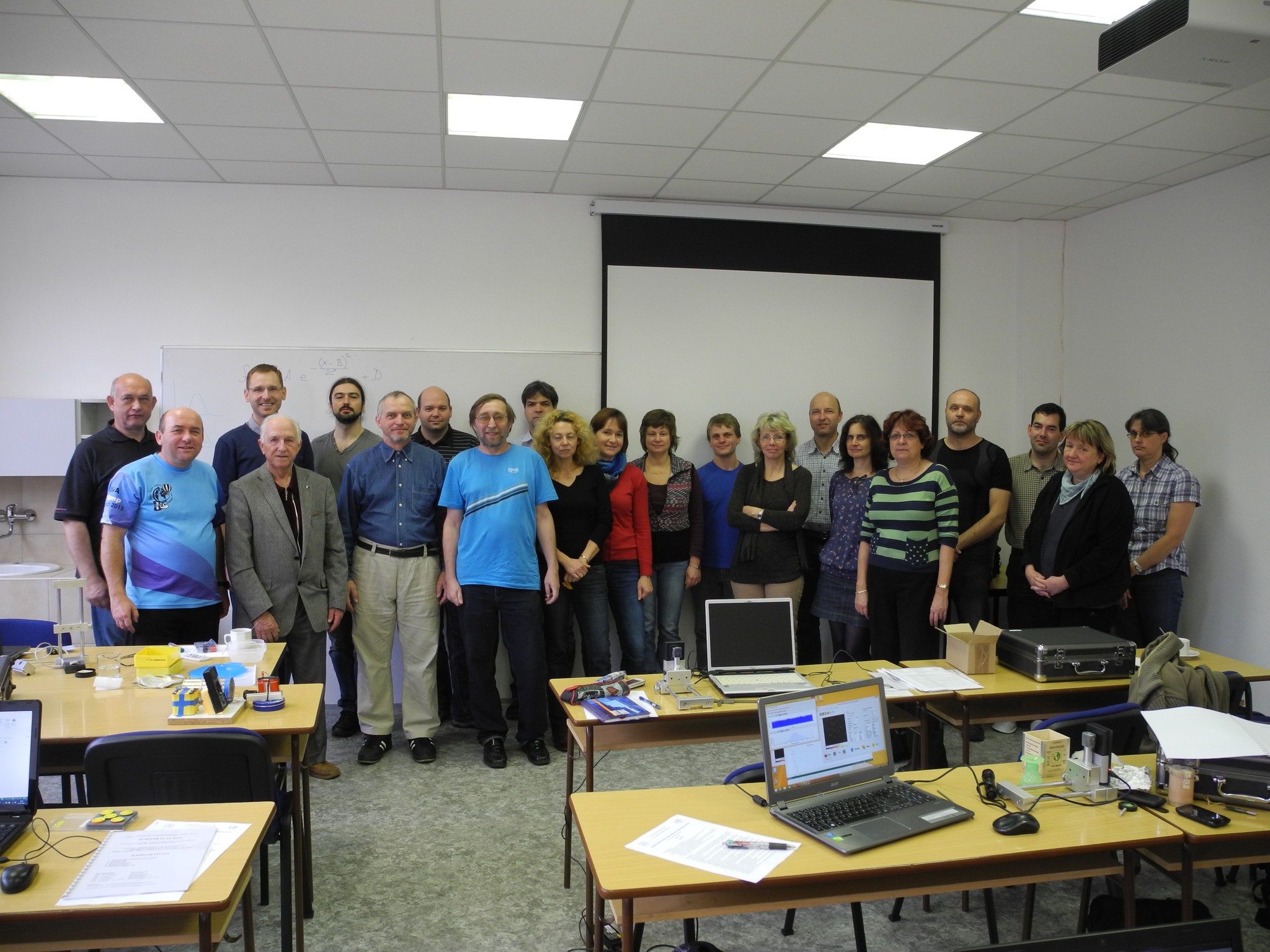 Progressive detection methods in atomic and particle physics education at middle and high school level
 NSS MIC IEEE Conference
 SEPnet, CERN@school Conference
 Lovci záhad - natáčení ČT ve spolupráci s ÚTEF
 Advanced detection methods in atomic and subatomic physics education.
 Listening to the universe by detection cosmic rays - visit of French and Czech students
 NSS MIC IEEE Conference
 15thIWORID
 NSS MIC IEEE Conference