Publication
> Articles in Impacted Journals
> 'Real-time X-ray microradiographic imaging and image correlation for local strain mapping in single trabecula under mechanical load'
Real-time X-ray microradiographic imaging and image correlation for local strain mapping in single trabecula under mechanical load
Author
| Doktor Tomáš | ITAM AS CR, v. v. i. |
| Jiroušek Ondřej, Doc. Ing. PhD. | Institute of Theoretical and Applied Mechanics, Academy of Sciences of the Czech Republic |
| Zlámal Petr, Ing. | ITAM AS CR, v. v. i. |
| Jandejsek Ivan, Ing. | IEAP |
Year
2011
Scientific journal
JINST 6 C11007 doi:10.1088/1748-0221/6/11/C11007
Web
Abstract
X-ray microradiography was employed to quantify the strains in loaded human trabecula. Samples of isolated trabeculae from human proximal femur were extracted and glued in a loading machine specially designed and manufactured for testing small specimens. The samples were then tested in tension and three-point bending until complete fracture of the specimen occured. To assess the deformation in the very small samples (thickness 100μm, length 1—2mm) a real-time microradiography in conjunction with digital image correlation (DIC) has been employed. Loaded samples were irradiated continuously by X-rays (Hamamatsu L8601-01 with 5μm spot) during the test. Radiographs were acquired using 0.25s exposure time with hybrid single-photon counting silicon pixel detector Medipix2. The distance between the source and detector was kept small to ensure radiographs of good quality for such a short exposure time. Design of the experimental loading device enables for precise control of the applied displacement which is important for the post-yield behavior assessment of trabeculae. Large dynamic range, high sensitivity and high contrast of the Medipix2 enables measuring even very small strains with DIC. Tested experimental setup enables to combine micromechanical testing of the basic building block of trabecular bone with time-lapse X-ray radiography to measure the strains and to assess the mechanical properties of single human trabecula as well as to capture the softening curve with sufficient precision.
Grants
| LC06041 | Research center "Fabrication, modification and characterization of materials by energetic radiation" |
| 103/09/2101 | Evaluation of the energy responsible for fracture advancing |
Projects
Cite article as:
T. Doktor, O. Jiroušek, P. Zlámal, I. Jandejsek, "Real-time X-ray microradiographic imaging and image correlation for local strain mapping in single trabecula under mechanical load", JINST 6 C11007 doi:10.1088/1748-0221/6/11/C11007 (2011)
Search
Recent events
Seattle, USA
8-15 Nov 2014
Surrey, United Kingdom
Sep. 8, 2014
April 24, 2014
3 Apr 2014
Seoul, Korea
27 Oct - 2 Nov 2013
Paris
23-27 June 2013
29 Oct - 3 Nov 2012


 Experimental physics
with polarized protons, neutrons and deuterons
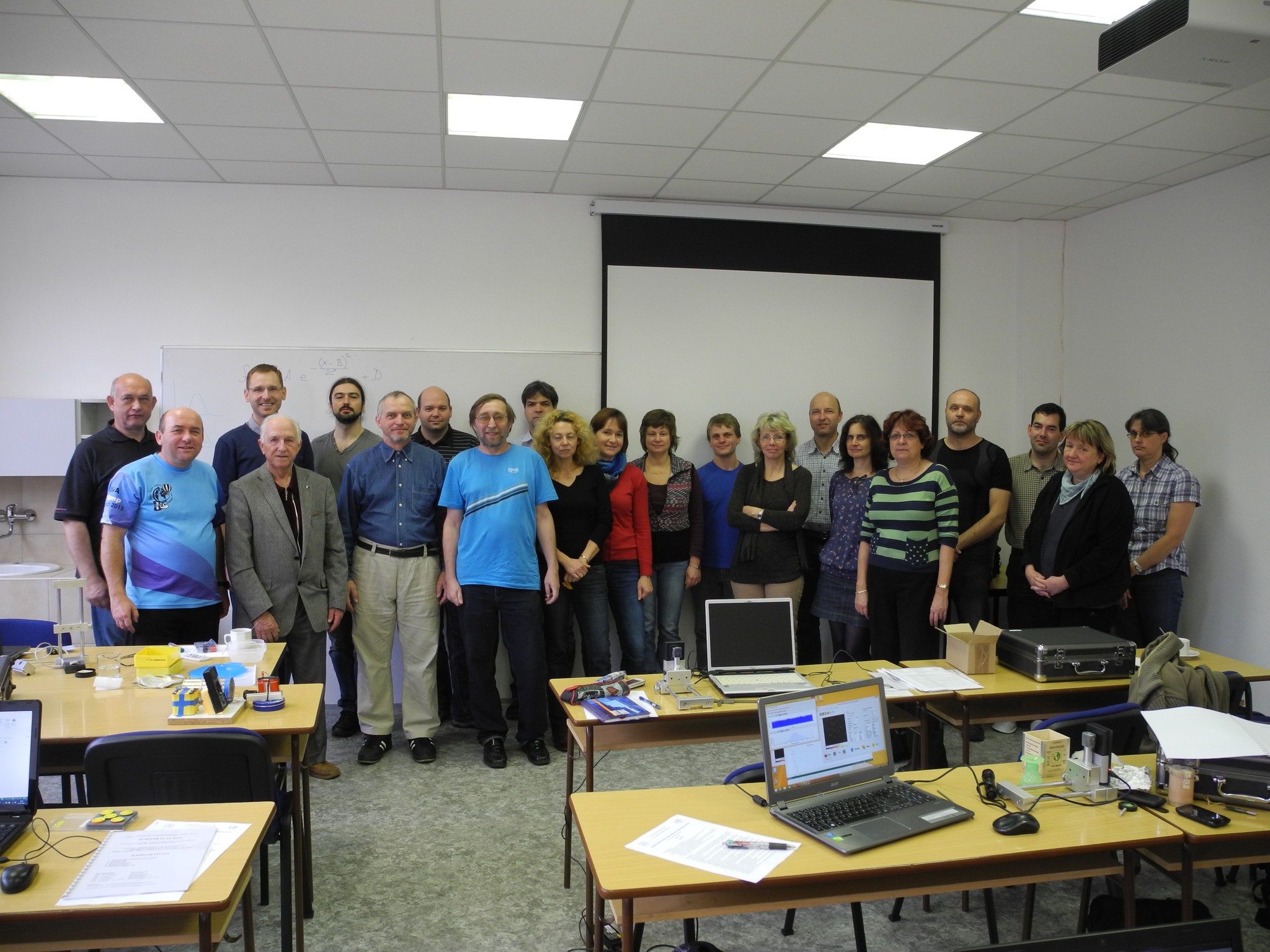 Progressive detection methods in atomic and particle physics education at middle and high school level
 NSS MIC IEEE Conference
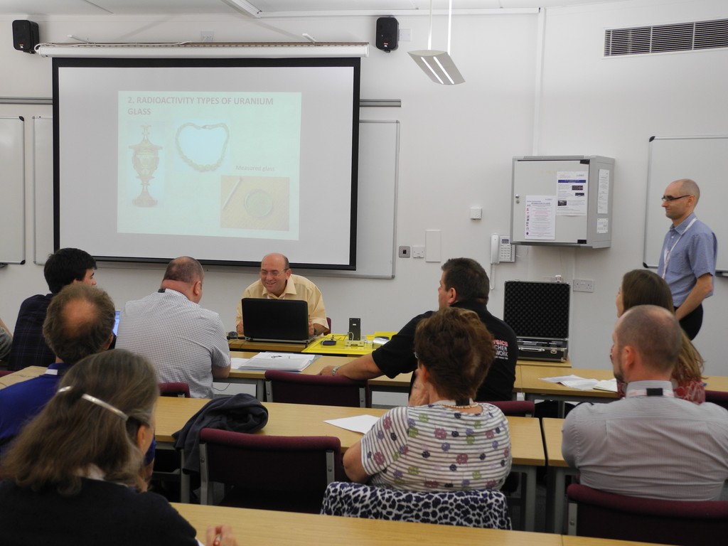 SEPnet, CERN@school Conference
 Lovci záhad - natáčení ČT ve spolupráci s ÚTEF
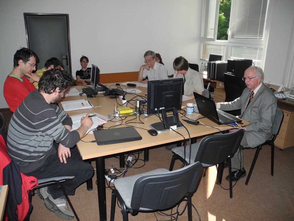 Advanced detection methods in atomic and subatomic physics education.
 Listening to the universe by detection cosmic rays - visit of French and Czech students
 NSS MIC IEEE Conference
 15thIWORID
 NSS MIC IEEE Conference