


Partial tasks
> Radiation Imaging in Material Science
Radiation Imaging in Material Science
Method “X Ray Dynamic Defectoscopy (XRDD)” uses Medipix device for the observation of time evolution of damage processes in a loaded specimen body. Even though the specimen is loaded monotonously and slowly, damaging can proceed discontinuously and relatively fast. Therefore high frame rate acquisition of X-ray images is crucial for real dynamic (time dependent) defectoscopy.
The method enables the observation of material density variations with micrometric accuracy. The test sample is illuminated by X rays during the loading process. Measured changes in transmission represent effective alterations in the sample thicknesses which are understood as weakening of the material due to damage volume fraction.
Sensitivity of the XRDD method depends on the ratio between incident and transmitted X ray beam intensity and on the fraction of scattered photons, the efficiency of the Medipix2 device at the X-ray energy used and the spectra of the X-ray tube.
Responsible person
Co-workers
Grants:
| Number | Name | Agency |
| LC06041 | Research center "Fabrication, modification and characterization of materials by energetic radiation" | MŠMT |
| 106/04/0567 | Study of damage zone in high ductile materials in vicinity of crack tip by X-Ray Dynamic Defectoscopy method | GAČR |
| MSM 6840770040 | Research Program 40: Usage of radionuclides and ionizing radiation. | MŠMT |
Articles in Impacted Journals
(21)
(21)
Table format
- J. Karch, B. Bartl, J. Dudák, J. Žemlička, F. Krejčí, "Non-destructive imaging of framents of historical beeswax seals using high-contrast X-ray micro-radiography and micro-tomography with large area photon-counting detector array", Micron (accepted) (2016)
- D. Vavřík, J. Jakůbek, I. Jandejsek, F. Krejčí, I. Kumpová, J. Žemlička, "Visualization of delamination in composite materials utilizing advanced X-ray imaging techniques", Journal of Instrumentation 10 C04012 (2015)
- F. Krejčí, M. Slavíková, J. Žemlička, J. Jakůbek, P. Kotlík, "High-contrast X-ray Radiography Using Hybrid Semiconductor Pixel Detectors with 1 mm thick Si sensor as a Tool for Monitoring Liquids in Natural Building", Journal of Instrumentation 9 C07014 (2014)
- M. Slavíková, F. Krejčí, J. Žemlička, M. Pech, P. Kotlík, J. Jakůbek, "X-ray Radiography and Tomography for Monitoring the Penetration Depth of Consolidants in Opuka - the Building stone of Prague Monuments ", Journal of Cultural Heritage 13 (4), 357-364 (2012)
- D. Vavřík, "CT Artefact Reduction by Signal to Thickness Calibration Function Shaping", NIM A, Volume 633, Supplement 1, May 2011, Pages S152-S155
- O. Jiroušek, I. Jandejsek, D. Vavřík, "Evaluation of strain field in microstructures using micro-CT and digital volume correlation", JINST 6 C01039 doi:10.1088/1748-0221/6/01/C01039 (2011)
- I. Jandejsek, P. Soukup, J. Jakůbek, "Image processing for X-ray transmission radiography with 3D voxel detector", JINST 6 C12061 doi:10.1088/1748-0221/6/12/C12061 (2011)
- D. Vavřík, P. Soukup, "Metal Grain Structure Resolved with Table-top micro-Tomographic System", JINST_007P_1011 (2011)
- T. Nguyen, D. Vavřík, E. Lehmann, I. Jeon, "Neutron Analysis for Microvoids in an Adhesive Layer between High X-ray Attenuation Materials", Appl. Phys. Express 4 (2011) 066401
- T. Doktor, O. Jiroušek, P. Zlámal, I. Jandejsek, "Real-time X-ray microradiographic imaging and image correlation for local strain mapping in single trabecula under mechanical load", JINST 6 C11007 doi:10.1088/1748-0221/6/11/C11007 (2011)
- U. Koester, C. Granja, J. Jakůbek, J. Uher, J. Vacík, "Slow-neutron-induced charged-particle emission-channeling-measurements with Medipix detectors", Nucl. Instr. Methods A 633 (2011) S267-S269
- P. Soukup, J. Jakůbek, I. Jandejsek, J. Žemlička, "X-ray color imaging with 3D sensitive voxel detector", JINST 6 C12014 doi:10.1088/1748-0221/6/12/C12014 (2011)
- I. Jandejsek, F. Nachtrab, N. Uhlmann, D. Vavřík, "X-ray dynamic defectoscopy utilizing digital image correlation", NIM A, Volume 633, Supplement 1, May 2011, Pages S185-S18
- D. Vavřík, J. Jakůbek, "Radiogram enhancement and linearization using the beam hardening correction method", NIM A, Vol. 607, Issue 1, p. 212-214 (2009)
- D. Vavřík, J. Jakůbek, T. Holý, "Micrometric scale measurement of material structure moving utilizing μ-radiographic technique", NIM A, Vol. 591, Issue 1, p. 24-27 (2008)
- J. Jakůbek, C. Granja, J. Dammer, R. Tykva, R. Hanus, J. Uher, Z. Vykydal, "Phase contrast enhanced high resolution X-ray imaging and tomography of soft tissue", Nucl. Instr. and Meth. A, Volume: 571, Issue: 1-2, Pages: 69-72, doi: 10.1016/j.nima.2006.10.031 (2007)
- J. Jakůbek, D. Vavřík, T. Holý, M. Jakůbek, Z. Vykydal, "Experimental system for high resolution X-ray transmission radiography", Nucl. Instr. and Meth. A, Volume: 563, Issue 1, Pages: 278-281, doi: 10.1016/j.nima.2006.01.033
- J. Jakůbek, D. Vavřík, S. Pospíšil, J. Uher, "Quality of X-ray transmission radiography based on single photon counting pixel device", NIM A Vol. 546, pages 113-117 (2005)
- J. Jakůbek, T. Holý, S. Pospíšil, D. Vavřík, "Tomography for XRDD", NIM A Vol. 531, pages 307-313 (2004)
- J. Jakůbek, S. Pospíšil, D. Vavřík, J. Visschers, "Resolution and stability tests of a Medipix-1 pixel detector for X-ray dynamic defectoscopy", NIM A Vol. 509, pages 294-301 (2003)
Search
Recent events
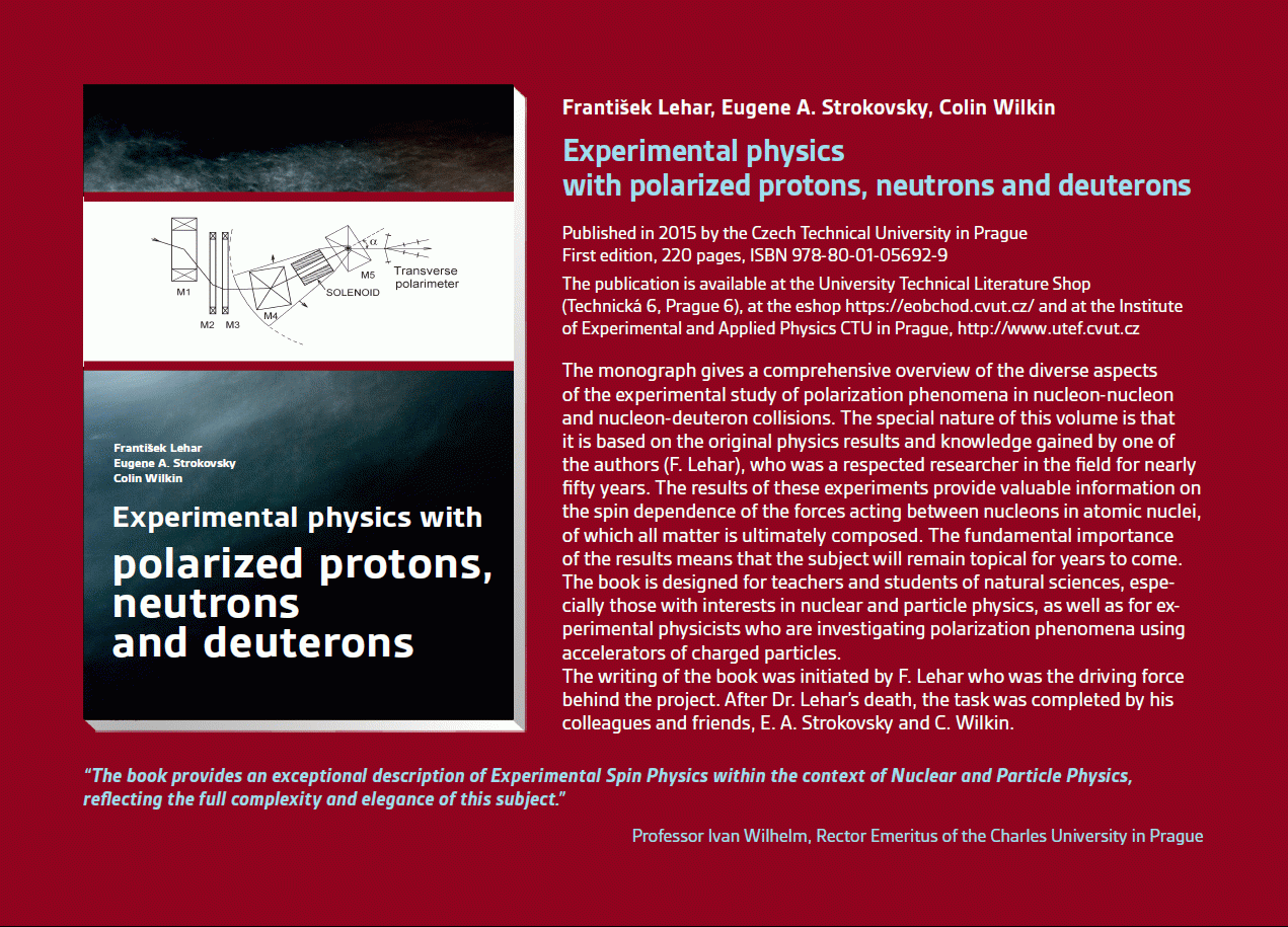 Experimental physics
with polarized protons, neutrons and deuterons
Experimental physics
with polarized protons, neutrons and deuterons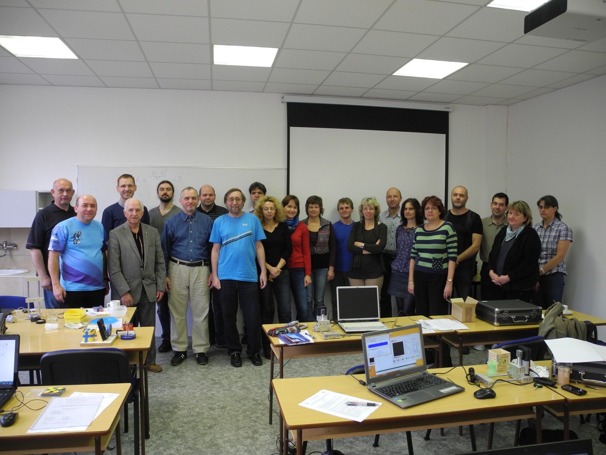 Progressive detection methods in atomic and particle physics education at middle and high school level
 NSS MIC IEEE Conference
Seattle, USA
8-15 Nov 2014
 SEPnet, CERN@school Conference
Surrey, United Kingdom
Sep. 8, 2014
 Lovci záhad - natáčení ČT ve spolupráci s ÚTEF
 Advanced detection methods in atomic and subatomic physics education.
April 24, 2014
 Listening to the universe by detection cosmic rays - visit of French and Czech students
3 Apr 2014
Seoul, Korea
27 Oct - 2 Nov 2013
 15thIWORID
Paris
23-27 June 2013
 NSS MIC IEEE Conference
29 Oct - 3 Nov 2012
