Publication
> Articles in Conference Proceedings
> 'Investigations of Secondary Ion Distributions in Carbon Ion Therapy Using the Timepix Detector'
Investigations of Secondary Ion Distributions in Carbon Ion Therapy Using the Timepix Detector
Author
| Gwosch Klauss | Medical Physics in Radiation Oncology, German Cancer Research Center (DKFZ), Im Neuenheimer Feld 280, D-69120 Heidelberg, Germany |
| Hartmann B. | DKFZ Heidelberg |
| Jakůbek Jan, Ing. Ph.D. | IEAP |
| Granja Carlos, Doc. Ing. Ph.D. | IEAP |
| Soukup Pavel, Ing. Ph.D. | IEAP |
| Jaekel Oliver | eHeidelberger Ionenstrahl-Therapiezentrum HIT |
| Martisikova M., Dr. | DKFZ Heidelberg |
Year
2012
Scientific journal
Med. Physics 39 (2012) 3614-3615
Web
Abstract
Purpose: Due to the high conformity of carbon ion therapy, unpredictable changes in the patient's geometry or deviations from the planned beam properties can result in changes of the dose distribution. PET has been used successfully to monitor the actual dose distribution in the patient. However, it suffers from biological washout processes and low detection efficiency. The purpose of this contribution is to investigate the potential of beam monitoring by detection of prompt secondary ions emerging from a homogeneous phantom, simulating a patient's head. Methods: Measurements were performed at the Heidelberg Ion‐Beam Therapy Center (Germany) using a carbon ion pencil beam irradiated on a cylindrical PMMA phantom (16cm diameter). For registration of the secondary ions, the Timepix detector was used. This pixelated silicon detector allows position‐resolved measurements of individual ions (256×256 pixels, 55μm pitch). To track the secondary ions we used several parallel detectors (3D voxel detector). Results: For monitoring of the beam in the phantom, we analyzed the directional distribution of the registered ions. This distribution shows a clear dependence on the initial beam energy, width and position. Detectable were range differences of 1.7mm, as well as vertical and horizontal shifts of the beam position by 1mm. To estimate the clinical potential of this method, we measured the yield of secondary ions emerging from the phantom for a beam energy of 226MeV/u. The differential distribution of secondary ions as a function of the angle from the beam axis for angles between 0 and 90° will be presented. In this setup the total yield in the forward hemisphere was found to be in the order of 10−1 secondary ions per primary carbon ion. Conclusions: The presented measurements show that tracking of secondary ions provides a promising method for non‐invasive monitoring of ion beam parameters for clinical relevant carbon ion fluences. Research with the pixel detectors was carried out in frame of the Medipix Collaboration.
Cite article as:
K. Gwosch, B. Hartmann, J. Jakůbek, C. Granja, P. Soukup, O. Jaekel, M. Martisikova, "Investigations of Secondary Ion Distributions in Carbon Ion Therapy Using the Timepix Detector", Med. Physics 39 (2012) 3614-3615 (2012)
Search
Recent events
Seattle, USA
8-15 Nov 2014
Surrey, United Kingdom
Sep. 8, 2014
April 24, 2014
3 Apr 2014
Seoul, Korea
27 Oct - 2 Nov 2013
Paris
23-27 June 2013
29 Oct - 3 Nov 2012


 Experimental physics
with polarized protons, neutrons and deuterons
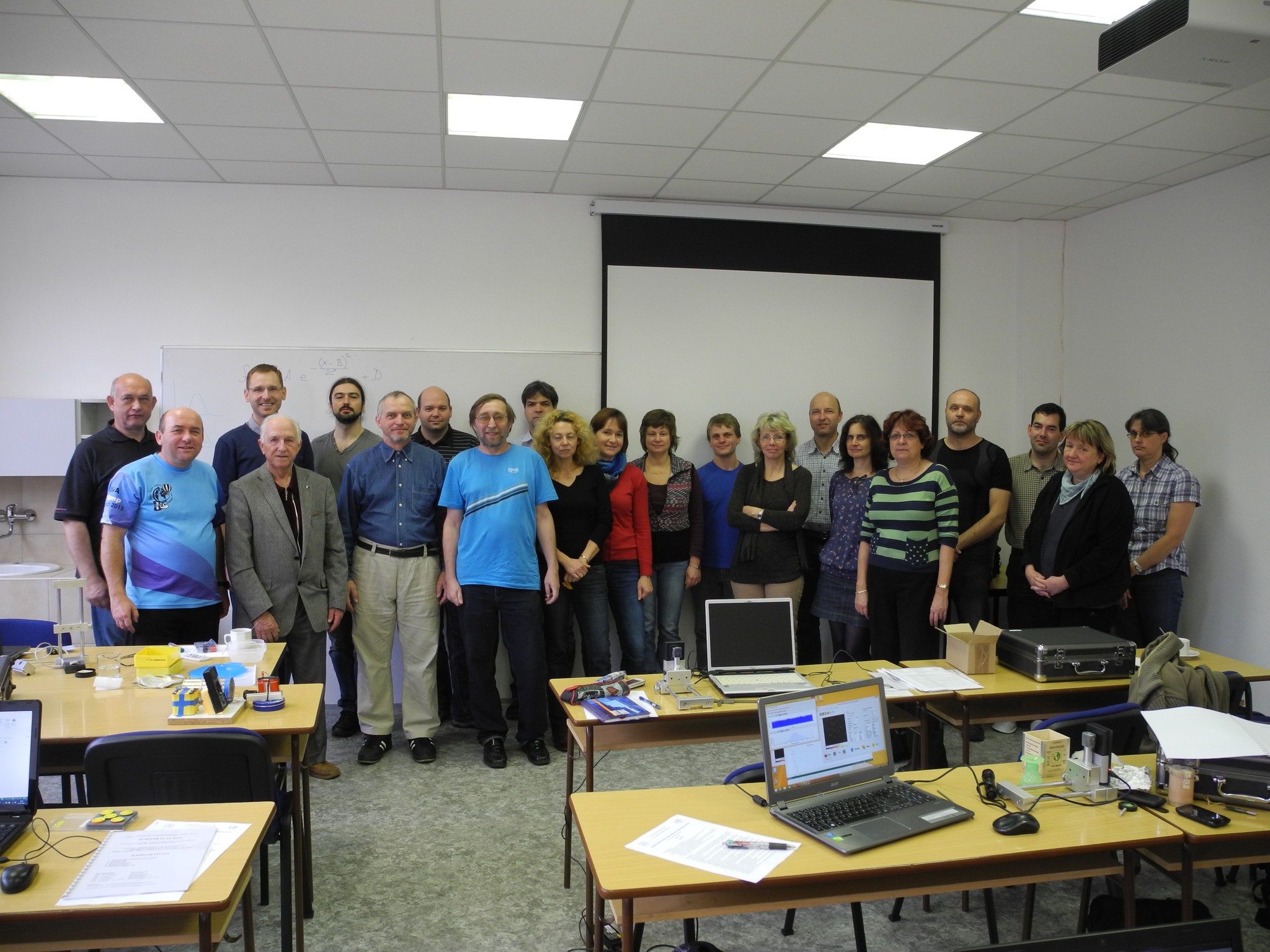 Progressive detection methods in atomic and particle physics education at middle and high school level
 NSS MIC IEEE Conference
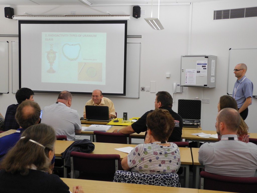 SEPnet, CERN@school Conference
 Lovci záhad - natáčení ČT ve spolupráci s ÚTEF
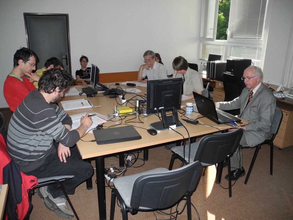 Advanced detection methods in atomic and subatomic physics education.
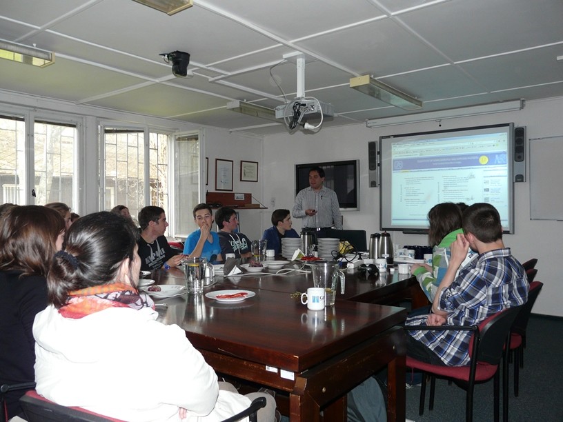 Listening to the universe by detection cosmic rays - visit of French and Czech students
 NSS MIC IEEE Conference
 15thIWORID
 NSS MIC IEEE Conference