Publikace
> Články v impaktovaných časopisech
> 'Non-invasive monitoring of therapeutic carbon ion beams in a homogeneous phantom by tracking of secondary ions '
Non-invasive monitoring of therapeutic carbon ion beams in a homogeneous phantom by tracking of secondary ions
Autor
| Gwosch Klauss | Medical Physics in Radiation Oncology, German Cancer Research Center (DKFZ), Im Neuenheimer Feld 280, D-69120 Heidelberg, Germany |
| Hartmann Bernadette | Department of Medical Physics in Radiation Oncology, German Cancer Research Center |
| Jakůbek Jan, Ing. Ph.D. | UTEF |
| Granja Carlos, Doc. Ing. Ph.D. | UTEF |
| Soukup Pavel, Ing. Ph.D. | UTEF |
| Martisikova Maria | Department of Medical Physics in Radiation Oncology, German Cancer Research Center |
| Jaekel Oliver | eHeidelberger Ionenstrahl-Therapiezentrum HIT |
Rok
2013
Časopis
Physics in Medicine and Biology, Vol. 58, p3755, doi:10.1088/0031-9155/58/11/3755
Web
Obsah
Radiotherapy with narrow scanned carbon ion beams enables a highly accurate treatment of tumours while sparing the surrounding healthy tissue. Changes in the patient's geometry can alter the actual ion range in tissue and result in unfavourable changes in the dose distribution. Consequently, it is desired to verify the actual beam delivery within the patient. Real-time and non-invasive measurement methods are preferable. Currently, the only technically feasible method to monitor the delivered dose distribution within the patient is based on tissue activation measurements by means of positron emission tomography (PET). An alternative monitoring method based on tracking of prompt secondary ions leaving a patient irradiated with carbon ion beams has been previously suggested. It is expected to help in overcoming the limitations of the PET-based technique like physiological washout of the beam induced activity, low signal and to allow for real-time measurements. In this paper, measurements of secondary charged particle tracks around a head-sized homogeneous PMMA phantom irradiated with pencil-like carbon ion beams are presented. The investigated energies and beam widths are within the therapeutically used range. The aim of the study is to deduce properties of the primary beam from the distribution of the secondary charged particles. Experiments were performed at the Heidelberg Ion Beam Therapy Center, Germany. The directions of secondary charged particles emerging from the PMMA phantom were measured using an arrangement of two parallel pixelated silicon detectors (Timepix). The distribution of the registered particle tracks was analysed to deduce its dependence on clinically important beam parameters: beam range, width and position. Distinct dependencies of the secondary particle tracks on the properties of the primary carbon ion beam were observed. In the particular experimental set-up used, beam range differences of 1.3 mm were detectable. In addition, variations in the beam width could be measured with a precision of 0.9 mm. Furthermore, shifts of the lateral beam position could be monitored with a sub-millimetre precision. The presented investigations demonstrate experimentally that the non-invasive measurement and analysis of secondary ion distributions around head-sized homogeneous objects provide information on the actual beam delivery. Beam range, width and position could be monitored with a precision attractive for therapeutic situations.
Granty
Projekty
Příklad citace článku:
K. Gwosch, B. Hartmann, J. Jakůbek, C. Granja, P. Soukup, M. Martisikova, O. Jaekel, "Non-invasive monitoring of therapeutic carbon ion beams in a homogeneous phantom by tracking of secondary ions ", Physics in Medicine and Biology, Vol. 58, p3755, doi:10.1088/0031-9155/58/11/3755 (2013)
Hledat
Události
21.-22. 11. 2014
Seattle, USA
8-15 Nov 2014
Surrey, Velká Británie
8. září 2014
9. září 2014
24. 4. 2014
3. 4. 2014
Seoul, Korea
27 Oct - 2 Nov 2013
Paris
23-27 June 2013
Anaheim, USA
29 Oct - 3 Nov 2012


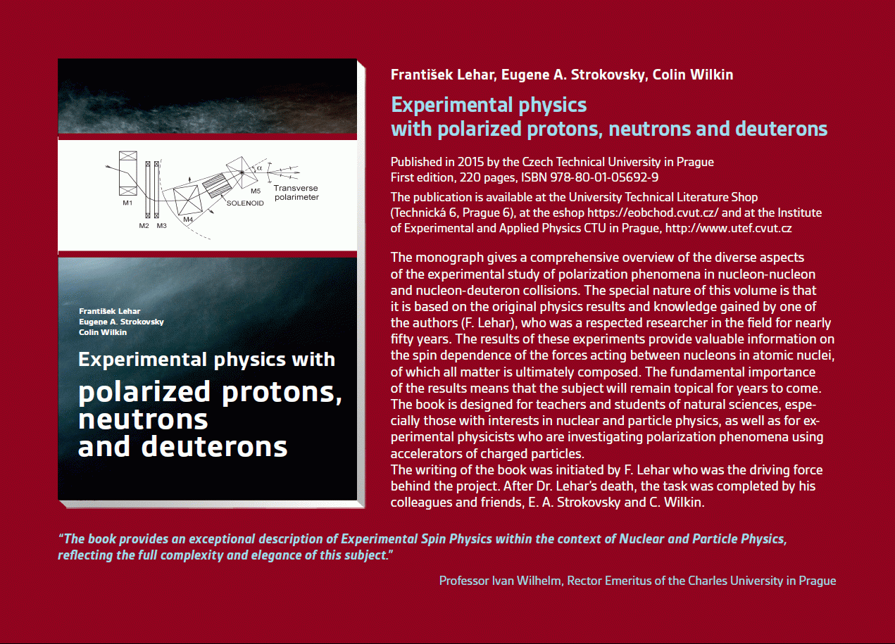 Experimental physics
with polarized protons, neutrons and deuterons
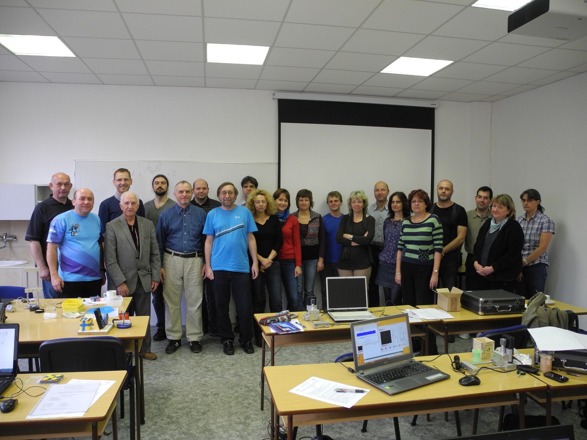 Progresivní detekční metody ve výuce subatomové a částicové fyziky
na ZŠ a SŠ
 NSS MIC IEEE Conference
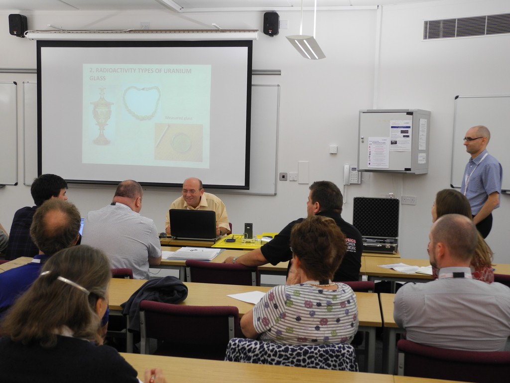 Konference SEPnet, CERN@school
 Lovci záhad - spolupráce ČT a ÚTEF
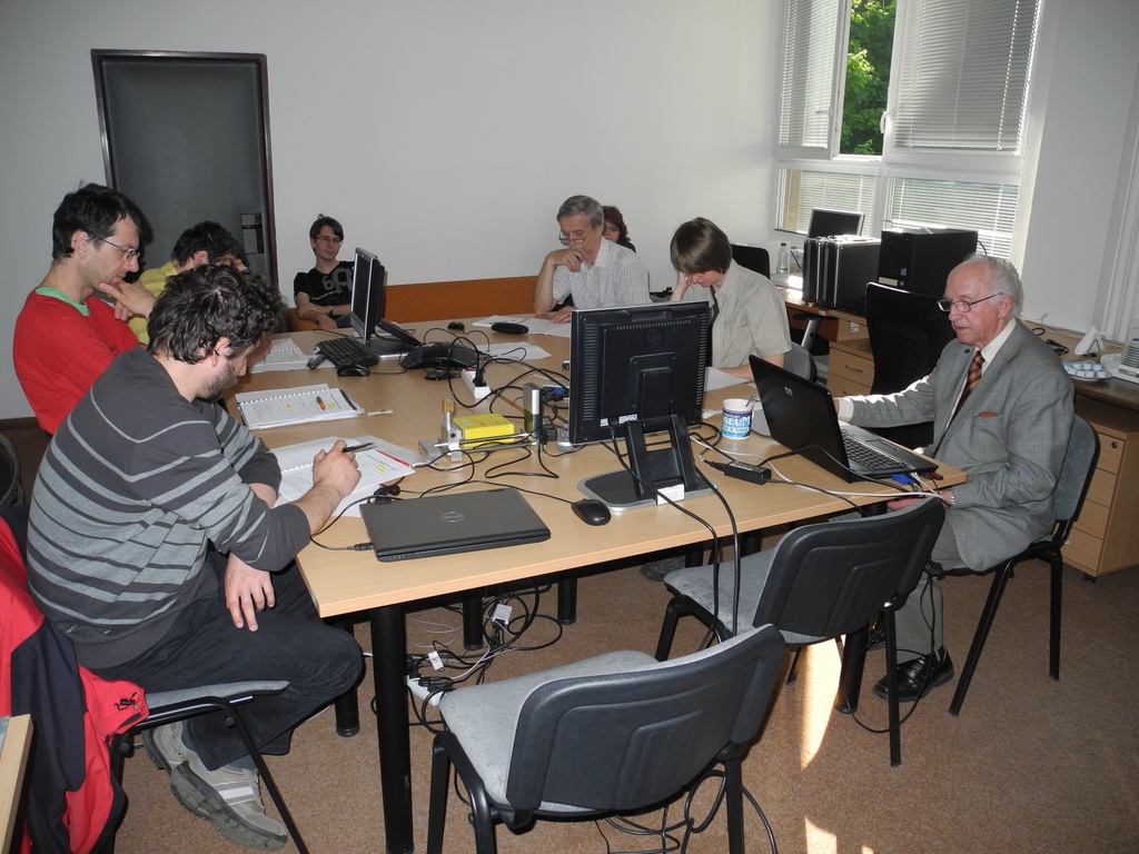 Progresivní detekční metody ve výuce subatomové a částicové fyziky na ZŠ a SŠ
 Návštěva v rámci projektu „Listening to the universe by detection cosmic rays“
 NSS MIC IEEE Conference
 15thIWORID
 NSS MIC IEEE Conference