
The nano/micro CT setup at the IEAP is dedicated to high-resolution investigation of internal structures of objects using X-rays. The system is built by the IEAP including hardware for the detector read-out, control software for the data acquisition, software for control of translation stages and CT reconstruction software.
X-ray source: The X-ray source used is a state-of-the-art FeinFocus® FXE 160.50 open-type tube with a dual head. A transmission sub-micro head is used when the application requires high spatial resolution which is reached using extremely high geometrical magnification in point-like source geometry (highest available spatial resolution ~ 700 nm without further postprocessing). The other head is a high-power oil-cooled direction tube with a conical tungsten anode and Aluminum or Beryllium output window. The higher output flux is available with spatial resolution about 6 µm. The mechanical stability of the system is provided by a shielded cabined embedded to the independent floor and thermal stabilization of the system. The open feature of the X-ray tube gives possibility to change the anode material together with the tube output material which enables optimization of the spectrum for a given application. Currently, following targets are available: W, W/Be, W/Diamond, Cu/Be, Mo/Be, Cr/Be, Cr/Be.
Detector equipment: The setup can be equipped by various detectors of the Medipix family (Medipix2, Medipix3, Timepix) which are available in the single chip configuration as well as the Quad device, stacked detector configurations and the large area hybrid pixel detector - 10×10 Timepix chips, available during 2014).
Translation stages: The system is equipped by 9 permanent translation and one rotation stages fully controlled by the Pixelman software; open architecture of the system enables with ease implementation of further stages (including software support, scripting tools etc.).
Applications: The non-destructive imaging in 2-D and 3-D (computed tomography) opens possibilities for interdisciplinary research in various fields such as biology (small animal imaging, for smaller samples including in vivo studies), material research (defectoscopy, material characterization), biomedicine (tissue characterization), culture heritage studies (artifacts visualization, stone consolidation studies) etc. Such projects are realized at the IEAP often with external collaborators. Besides X-ray radiography and CT-applications, due to a great variability of the installed equipment, the setup is often utilized for instrumentation and development of new imaging methods based on X-rays and hybrid semiconductor pixel detectors. From these techniques, phase contrast X-ray imaging, energy sensitive (color) X-ray radiography and tomography and fluorescent X-ray radiography are at the IEAP intensively studied.
Staff: Frantisek Krejci (contact person, beam time allowance), Jan Zemlicka (contact person), Jan Jakubek (head of the group), Jan Dudak, Ivan Jandejsek, Daniel Vavrik, Carla Palma, Kevin Loo. The system is placed in the main IEAP building Horska 3a/22 Prague. For proposals of scientific collaboration based on this system contact please us.





 Experimental physics
with polarized protons, neutrons and deuterons
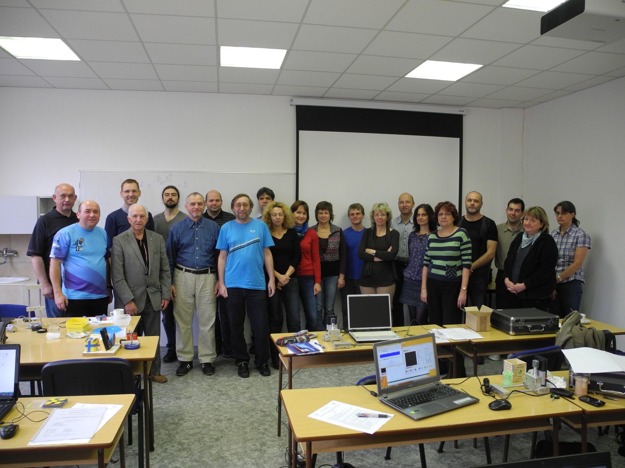 Progresivní detekční metody ve výuce subatomové a částicové fyziky
na ZŠ a SŠ
 NSS MIC IEEE Conference
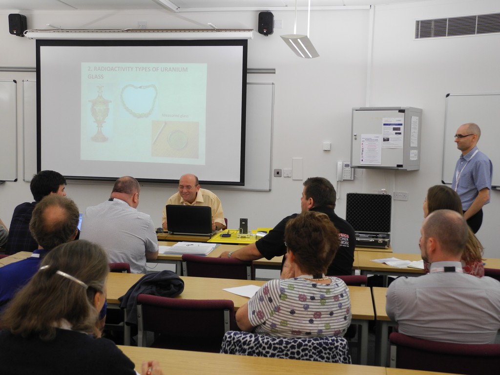 Konference SEPnet, CERN@school
 Lovci záhad - spolupráce ČT a ÚTEF
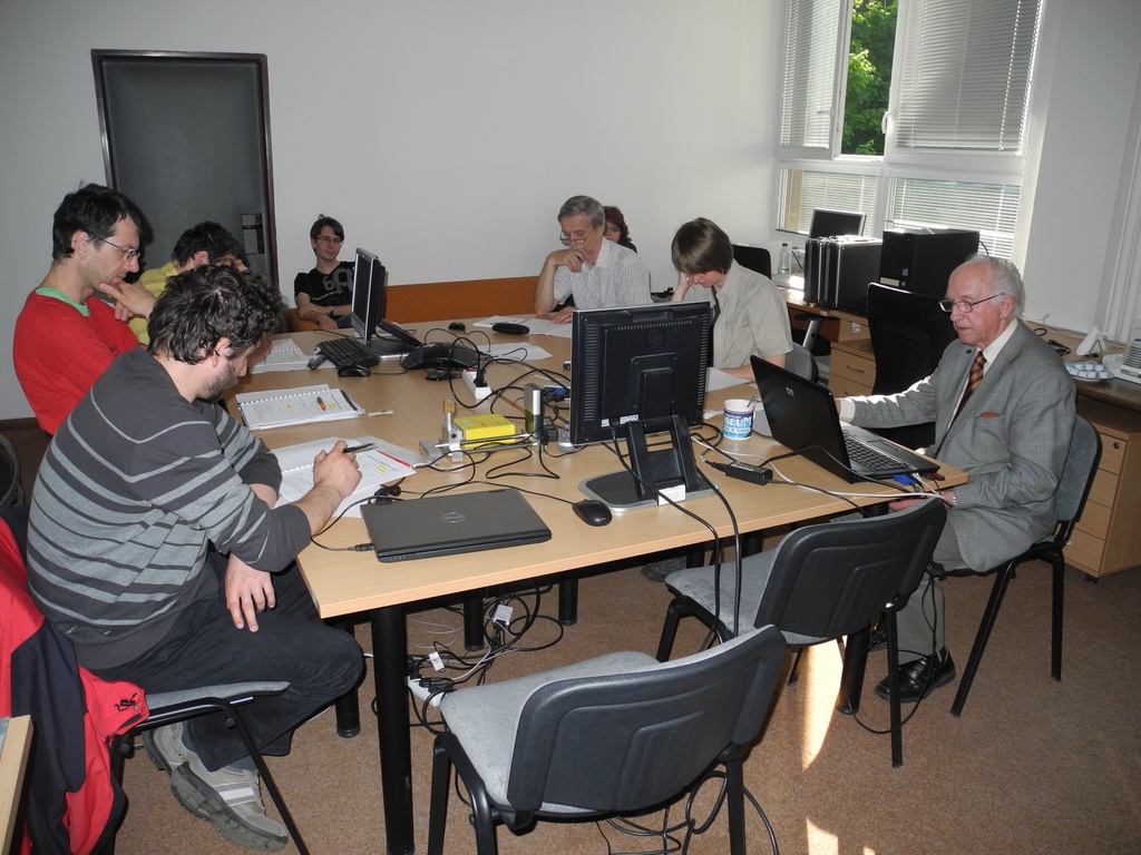 Progresivní detekční metody ve výuce subatomové a částicové fyziky na ZŠ a SŠ
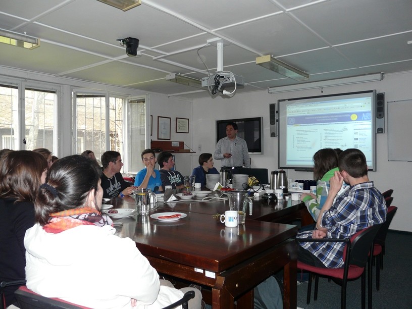 Návštěva v rámci projektu „Listening to the universe by detection cosmic rays“
 NSS MIC IEEE Conference
 15thIWORID
 NSS MIC IEEE Conference