Publikace
> Články v impaktovaných časopisech
> 'Microradiography of biological samples with Timepix'
Microradiography of biological samples with Timepix
Autor
| Dammer Jiří, Mgr. | UTEF |
| Weyda František, doc. RNDr. CSc. | Biologické centrum Akademie věd České republiky, v. v. i. |
| Benes J. | Charles University in Prague, First Faculty of Medicine |
| Sopko Vitek, Ing., Ph.D. | UTEF |
| Jakůbek Jan, Ing. Ph.D. | UTEF |
| Vondracek V. | University Hospital Na Bulovce, Department of Radiological Physics, Prague, Czech Republic |
Rok
2011
Časopis
JINST 6 C11005 doi:10.1088/1748-0221/6/11/C11005
Web
Obsah
Microradiography is an imaging technique using X-rays in the study of internal structures of objects. This rapid and convenient imaging tool is based on differential X-ray attenuation by various tissues and structures within the biological sample. The non-absorbed radiation is detected with a suitable detector and creates a radiographic image. In order to detect the differential properties of X-rays passing through structures sample with different compositions, an adequate high-quality imaging detector is needed. We describe the recently developed radiographic apparatus, equipped with Timepix semiconductor pixel detector. The detector is used as an imager that counts individual photons of ionizing radiation, emitted by an X-ray tube FeinFocus with tungsten, copper or molybdenum anode. Thanks to the wide dynamic range, time over threshold mode — counter is used as Wilkinson type ADC allowing direct energy measurement in each pixel of Timepix detector and its high spatial resolution better than 1μm, the setup is particularly suitable for radiographic imaging of small biological samples. We are able to visualize some internal biological processes and also to resolve the details of insects (morphology) using different anodes. These anodes generate different energy spectra. These spectra depend on the anode material. The resulting radiographic images varies according to the selected anode. Tiny live insects are an ideal object for our studies.
Granty
Projekty
Medipix
Zobrazování biologických vzorků pomocí rentgenového záření
Rentgenová absorpční a fázová radiografie a tomografie
Zobrazování biologických vzorků pomocí rentgenového záření
Rentgenová absorpční a fázová radiografie a tomografie
Ocenění
Award for the first best poster
Příklad citace článku:
J. Dammer, F. Weyda, J. Benes, V. Sopko, J. Jakůbek, V. Vondracek, "Microradiography of biological samples with Timepix", JINST 6 C11005 doi:10.1088/1748-0221/6/11/C11005 (2011)
Hledat
Události
21.-22. 11. 2014
Seattle, USA
8-15 Nov 2014
Surrey, Velká Británie
8. září 2014
9. září 2014
24. 4. 2014
3. 4. 2014
Seoul, Korea
27 Oct - 2 Nov 2013
Paris
23-27 June 2013
Anaheim, USA
29 Oct - 3 Nov 2012


 Experimental physics
with polarized protons, neutrons and deuterons
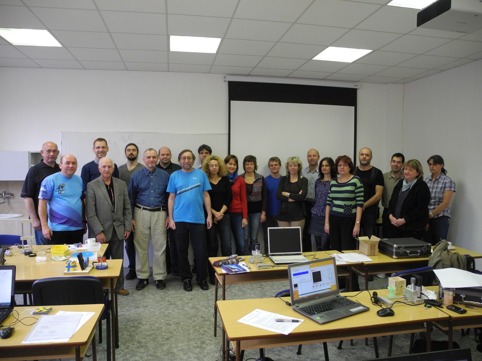 Progresivní detekční metody ve výuce subatomové a částicové fyziky
na ZŠ a SŠ
 NSS MIC IEEE Conference
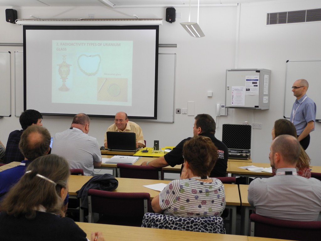 Konference SEPnet, CERN@school
 Lovci záhad - spolupráce ČT a ÚTEF
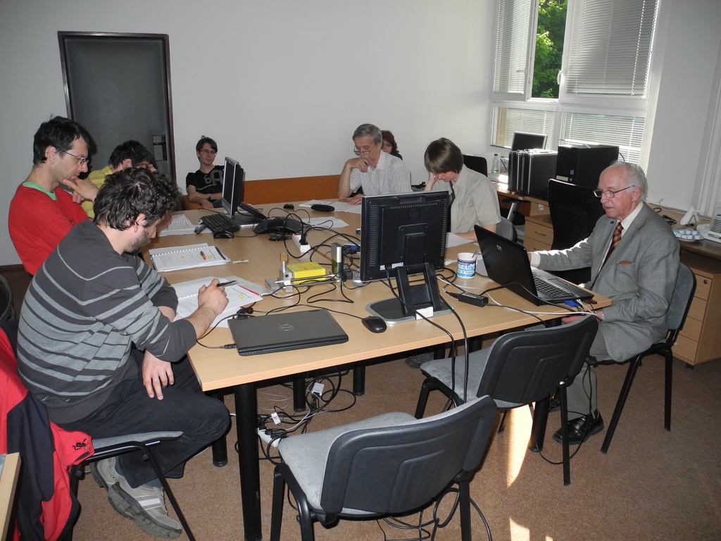 Progresivní detekční metody ve výuce subatomové a částicové fyziky na ZŠ a SŠ
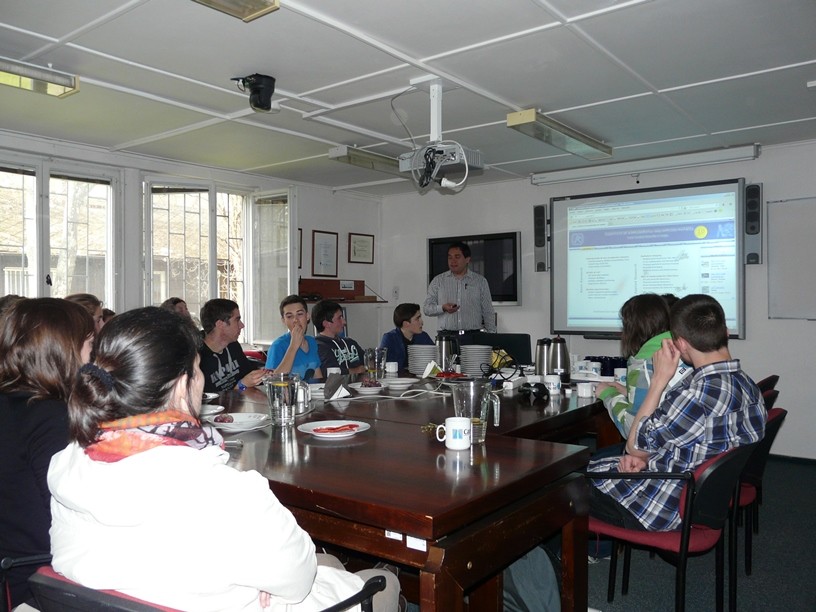 Návštěva v rámci projektu „Listening to the universe by detection cosmic rays“
 NSS MIC IEEE Conference
 15thIWORID
 NSS MIC IEEE Conference